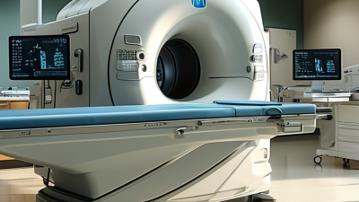
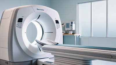
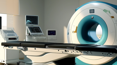
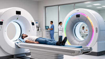
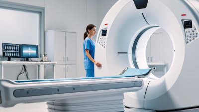
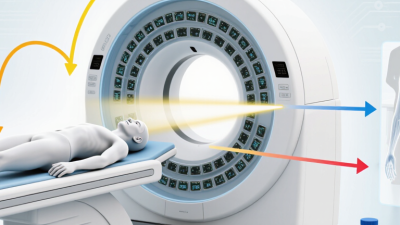
The leading Ct Equipment types in 2026 are not defined by one universal ranking. The right choice depends on the exam, patient population, clinical setting, and available budget. A busy hospital may prioritize a multidetector CT scanner that handles routine and emergency imaging. A cardiac center may focus on fast image acquisition and motion management. Different needs, different machines.
This guide compares conventional and multidetector systems, cardiac CT, dual-energy and spectral scanners, cone-beam CT, mobile units, and photon-counting technology. It explains what each type is designed to do, where its advantages may matter, and what buyers should check beyond headline specifications. Look closely at detector configuration, bore size, dose-management features, reconstruction tools, room requirements, and service support. Small details count. A wide bore, for example, can make positioning easier for some patients, while a compact room may limit installation options. More advanced features do not automatically mean better clinical value.
Technology also changes at an uneven pace. Photon-counting systems attract attention, but availability, cost, and suitability vary by market and use case. Rankings can simplify a complicated decision—and sometimes mislead. This overview therefore treats “top” as a practical comparison, not a claim that one scanner fits every facility. Clinical teams and imaging specialists should verify performance claims against their own protocols and requirements. Some trade-offs remain difficult to capture in a short comparison. That is worth remembering.
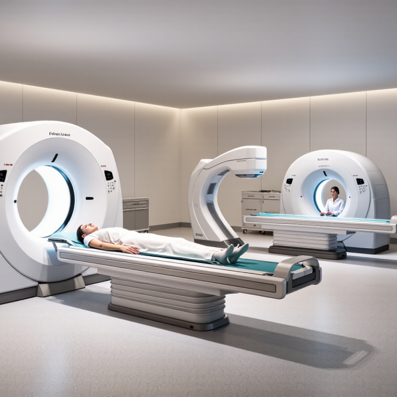
Multidetector CT scanners use multiple detector rows to capture data during each rotation. Systems with 16 to 320 rows cover many routine clinical needs, from chest imaging to cardiac studies. More rows can support wider coverage and faster acquisition, which helps when patients cannot hold their breath for long. But detector count alone does not determine image quality. Gantry rotation speed, reconstruction methods, protocol settings, and patient positioning all matter.
The numbers can mislead. A higher row count does not automatically mean lower radiation dose or better results for every exam. For example, a broad detector array may capture the heart in fewer beats, while a carefully adjusted protocol can be more important for a standard abdominal scan. The right system depends on the facility’s case mix, workflow, and staff expertise. Even experienced teams need to review protocols as technology and clinical needs change.

Dual-source CT uses two tube–detector pairs positioned around the patient. They collect data from different angles at the same time, helping capture a moving heart with less motion blur. In practical terms, this can support coronary imaging when a heartbeat leaves only a narrow window for clear measurements. Fast does not mean effortless.
A technical performance study by Flohr and colleagues reported 83 ms temporal resolution for an early dual-source CT system (European Radiology, 2006; 16:256–268). That figure describes the system tested, not every scanner available today. Actual image quality also depends on heart rhythm, scan protocol, patient size, and staff decisions. Motion still matters. Nor does dual-source design automatically mean lower radiation exposure; dose depends on how the examination is planned and performed.
Tips: Review the patient’s rhythm and clinical question before choosing ECG gating and scan settings. Keep the scan range focused, and check dose indicators after the exam. If the rhythm is irregular, image quality may still suffer; that limitation deserves honest discussion.
What Are the Top CT Equipment Types in 2026?
Wide-detector CT: 16 cm coverage can capture the heart in one rotation
A wide-detector CT system covers up to 16 cm along the patient’s body during one rotation. Because the heart can fit within this coverage in many adults, the scanner may acquire cardiac images without moving the table mid-scan. That can help limit motion-related image blur. The benefit is especially useful when assessing structures that move with each heartbeat. But it is not a guarantee of perfect images. Heart rhythm, patient size, breathing, and scan setup all matter.
Tips: Check the detector’s actual coverage and the clinical protocol, not just the headline specification. Ask how staff manage breath-holding and irregular heart rhythms. Small details matter.
One rotation sounds simple. The clinical picture is not. A wide detector can support rapid cardiac imaging, but image quality still depends on timing, dose settings, and appropriate patient preparation. It is easy to overstate the advantage: 16 cm coverage does not mean every heart scan is motion-free or suitable for every patient. The technology is most valuable when its speed matches a clear diagnostic need. Hospitals should also consider workflow, staff training, and how often cardiac studies are performed.
Photon-counting CT became a clinical reality in the United States when the FDA cleared the first system in September 2021, according to its 510(k) database. Unlike conventional detectors, which combine incoming X-rays into one signal, photon-counting detectors register individual photons and sort them by energy. That gives radiologists more information from each scan. It is a meaningful engineering shift, not just a sharper picture.
Technical reports in Radiology describe ultra-high-resolution image sections as fine as 0.2 millimeters on early photon-counting systems. That detail can help reveal tiny structures, such as small lung nodules or narrow coronary arteries. Published studies also report potential for lower radiation exposure in selected imaging protocols, but results depend on the body part, scan settings, and patient. The numbers are promising, not a universal guarantee. Real-world use still requires careful protocol design, staff training, and comparison with established scanners. A smaller pixel does not automatically mean a better diagnosis. FDA clearance marked an important milestone; it did not settle every question about cost, workflow, or long-term clinical benefit.
A practical overview of major CT system types, their technology, common uses, and distinguishing features.
| CT Equipment Type | How It Works | Common Uses | Key Characteristics | 2026 Context |
|---|---|---|---|---|
| Conventional Energy-Integrating Detector CT | Detector elements convert x-ray energy into an electrical signal, which is used to reconstruct cross-sectional images. | Routine head, chest, abdominal, pelvic, vascular, and musculoskeletal imaging. | A widely established CT design with broad clinical utility. Image quality and dose depend on the scanner, protocol, and patient factors. | Remains a major part of clinical CT practice across hospitals and imaging centers. |
| Photon-Counting CT | Specialized detectors register individual x-ray photons and measure their energies, enabling energy-resolved image data. | Diagnostic imaging applications where fine detail or spectral information may be useful, subject to clinical indication and local availability. | Can provide spectral information from a single acquisition and may support high spatial resolution. Performance and dose depend on the system and protocol. | The FDA cleared the first photon-counting CT system in 2021. This is a detector technology, not a replacement for every CT application. |
| Dual-Energy / Spectral CT | Acquires data at two or more x-ray energy levels using approaches such as dual-source scanning, rapid voltage switching, or layered detectors. | Material characterization, iodine assessment, virtual non-contrast imaging, and selected vascular, oncology, and musculoskeletal studies. | Produces energy-dependent information that can help distinguish materials; available features vary by scanner design. | A capability found in multiple CT system designs; spectral CT is a functional category and can overlap with other equipment types. |
| Wide-Detector CT | Uses a broad detector array along the patient axis to cover a larger anatomical region during each gantry rotation. | Cardiac imaging, dynamic studies, perfusion imaging, and examinations that benefit from broad anatomic coverage. | Can capture a larger volume in fewer rotations. The achievable coverage and temporal performance depend on the scanner configuration. | Used where its coverage and workflow advantages match clinical needs; it is not a separate imaging principle from conventional CT. |
| Mobile / Intraoperative CT | A CT scanner designed to be brought to or used within settings such as operating rooms, neurocritical care units, or procedural areas. | Selected neurosurgical procedures, intraoperative assessment, and imaging of patients who are difficult to transport. | Supports imaging near the point of care, but requires suitable space, workflow planning, and radiation-safety procedures. | A setting- and workflow-oriented category; available configurations and clinical uses vary by facility. |
| Cone-Beam CT (CBCT) | A cone-shaped x-ray beam and flat-panel detector rotate around the patient to produce three-dimensional images. | Dental and maxillofacial imaging, selected ENT applications, and image guidance in some procedural settings. | Can provide detailed images of bone and teeth. Soft-tissue contrast is generally more limited than in conventional medical CT. | Common in specialized imaging environments; its field of view and diagnostic suitability depend on the application and device. |
Note: These categories can overlap—for example, a system may offer spectral imaging and wide detector coverage. The most suitable equipment depends on the clinical task, patient population, workflow, and local regulatory clearance.
Cone-beam CT, often called CBCT, brings three-dimensional imaging into dental rooms and specialty clinics. Its compact design can fit spaces that cannot accommodate a conventional medical CT scanner. During a scan, the unit rotates around the patient’s head and captures many projections. Software combines them into a detailed volume of the teeth, jaw, and nearby structures.
That view can help clinicians assess impacted teeth, plan implant placement, and examine complex root or jaw anatomy. It can also support image-guided procedures by showing landmarks from several angles. A small field of view may be enough for one tooth; broader coverage may be needed for larger questions. The scan should match the clinical need. More images are not automatically better.
Positioning matters. The patient must remain still, and even slight movement can blur fine detail. Metal restorations may create streaks or distortions. The image can look impressively crisp, yet clarity is not the same as certainty. CBCT findings need interpretation alongside symptoms, examination, and other records. Radiation exposure varies with the device and settings, so clinicians should choose protocols carefully and avoid repeat scans when existing images answer the question. Small systems are useful, but they are not a shortcut around sound clinical judgment.
Cone-beam CT uses different field-of-view sizes for different clinical needs. These representative heights are approximate; actual scan dimensions vary by system and protocol.