Choosing the top CT imaging systems in 2026 requires more than comparing detector rows or promotional claims. The strongest systems must produce clear images, support dependable clinical decisions, and manage radiation responsibly. They should also fit real hospital conditions, including busy emergency departments, limited staffing, and demanding service schedules.
This guide examines leading CT imaging platforms through practical criteria. These include spatial resolution, scan speed, spectral imaging, photon-counting capability, dose efficiency, reconstruction software, workflow integration, and long-term operating costs. A scanner may deliver impressive images on a laboratory brochure yet perform less effectively when patient movement, metal implants, or high daily volume becomes involved.
“The future of radiology is 3D,” says Dr. Elliot K. Fishman, a recognized authority in three-dimensional imaging and radiology. His observation remains relevant in 2026, as advanced visualization and quantitative analysis increasingly influence diagnosis and treatment planning. Still, technology is not the whole story. Skilled technologists and experienced radiologists matter greatly.
Not every hospital needs the most expensive platform. Sometimes, a reliable mid-range system provides better value. That may be an uncomfortable conclusion.
This overview compares major CT imaging systems without treating one manufacturer as universally superior. It considers image quality, patient safety, workflow, maintenance, and clinical flexibility. Evidence can also be uneven, because product updates and regional configurations change quickly. Readers should verify current specifications, regulatory clearances, and independent performance data before making a purchasing decision. The best system is not always the newest. It is the one that consistently serves patients, clinicians, and the institution’s actual needs.
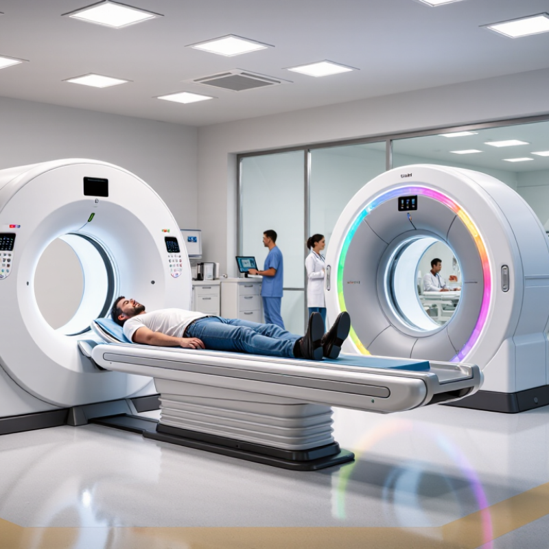
A leading CT imaging system in 2026 is defined by balanced clinical performance, not impressive specifications alone. It should produce sharp images at appropriate radiation doses, even when patients move or cannot hold their breath. Fast acquisition matters, but consistency matters more. A scan completed quickly is not useful if artifacts force a repeat examination.
Radiographers need clear protocols, responsive controls, and patient-centered workflow design. A well-designed system can automatically adjust exposure to body size and anatomy. It can also support reconstruction tools that reveal subtle findings without creating distracting visual noise. Artificial intelligence may assist with positioning, image review, and prioritization, but qualified professionals must remain responsible for clinical decisions. That boundary protects both patients and diagnostic quality.
Reliability is equally important. Hospitals should examine uptime records, service response times, cybersecurity practices, and compatibility with existing information systems. Independent clinical evidence is more persuasive than promotional claims. Real-world testing should include emergency cases, pediatric examinations, and patients with metal implants. Small weaknesses appear there. Even experienced teams can overvalue speed or automation. A leading system should make errors easier to detect, not easier to overlook. Its interface must explain alerts clearly, because vague warnings waste time in a crowded control room. Regular staff feedback can reveal problems that laboratory testing misses. Performance also depends on training, maintenance, and thoughtful protocol review.
Clinical performance is not judged by speed alone. A strong CT system must produce diagnostic images with controlled radiation exposure. The American College of Radiology’s Dose Index Registry shows substantial dose variation among facilities performing similar examinations. That gap suggests protocol quality matters as much as hardware.
Evaluation should include low-contrast detectability, spatial resolution, motion control, reconstruction accuracy, and workflow reliability. The European Commission’s 2024 radiation-protection guidance also emphasizes justification and optimization for every CT examination. Image quality must fit the clinical question. A sharper image is not always a better image. Sometimes, it only adds noise or dose.
Tips: Compare systems using identical phantom tests and patient protocols. Review CTDIvol and DLP against local diagnostic reference levels. Check pediatric and emergency presets separately. Ask how often protocols are audited. The IAEA recommends regular quality-control testing, including image uniformity, noise, and dose verification. These checks expose weaknesses that a polished demonstration can hide. Operator experience also changes results. That part is easy to underestimate. A technically advanced scanner can still perform poorly when protocols are outdated or staff training is inconsistent. Any ranking should therefore combine published performance data, service records, clinical feedback, and independent acceptance testing.
What Are the Top CT Imaging Systems in 2026?
The leading CT imaging systems are defined by clinical purpose, not by one universal ranking. High-end platforms may combine wide-area detectors, dual-energy imaging, and advanced iterative reconstruction. These features can improve tissue characterization while controlling radiation exposure. Some systems also offer photon-counting technology, which captures more detailed spectral information. This may help clinicians assess small vessels, lung nodules, or complex bone structures. Performance still depends on protocol design, patient movement, and staff experience.
Cardiac-focused systems often emphasize rapid rotation and motion correction. They can produce clearer coronary images during short breath-holds. Emergency departments may prefer fast table movement and automated positioning. Oncology teams may value consistent low-dose follow-up scans and reliable contrast timing. Artificial intelligence can support reconstruction and workflow, but it does not replace clinical judgment. In practice, a technically advanced scanner may underperform when protocols are poorly adjusted. That is an easy detail to overlook.
Tips: Compare systems by real clinical workload. Ask for measured dose data, not only brochure claims. Review image quality using local cases. Check service coverage, staff training, software updates, and compatibility with existing systems. A trial scan can reveal practical problems, including noise, delays, or difficult patient positioning. Independent evaluation remains important. No system fits every hospital perfectly.
Major CT imaging system classes and their distinctive features
How to read the chart: Detector coverage indicates the approximate anatomical length captured in one gantry rotation. Wider coverage can improve volumetric imaging speed and support dynamic examinations, while higher detector-row systems are commonly used for routine, cardiac, and advanced spectral applications.
Values are representative technical figures for current CT system classes and may vary by configuration, acquisition protocol, and reconstruction method.
What Are the Top CT Imaging Systems in 2026?
Clinical Uses Across Diagnostic and Specialized Imaging
The strongest CT systems in 2026 are judged by clinical usefulness, not speed alone. Emergency departments need rapid head and body scans for stroke, trauma, and internal bleeding. Oncology teams require thin-slice images, contrast timing, and dependable lesion measurements. The International Agency for Research on Cancer reported 20 million new cancer cases worldwide in 2022. That burden makes reproducible CT imaging essential. Still, no scanner is perfect.
Dual-energy and photon-counting CT support material separation, vascular assessment, and tissue characterization. These features can clarify kidney stones, pulmonary embolism, gout, and complex cardiac anatomy. The American College of Radiology’s appropriateness guidance continues to emphasize selecting imaging according to the clinical question. Radiation control also matters. The IAEA states that justification and optimization should guide every medical exposure. Modern systems may reduce dose, but protocol design remains decisive. Hardware cannot correct poor positioning or weak clinical reasoning.
Specialized CT is also expanding in surgical planning and image-guided intervention. Spectral maps can reveal iodine distribution, while high-resolution acquisitions may improve temporal bone and lung evaluation. Reports from the OECD show persistent differences in diagnostic equipment access between health systems. That gap affects outcomes more than specifications alone. I may be too optimistic about automation; radiologist oversight remains necessary, especially with unusual anatomy, motion, or misleading artifacts.
| CT System Category | Typical Detector Configuration | Primary Clinical Uses | Specialized Imaging Applications | Key Clinical Advantages | Important Limitations | Best-Fit Care Setting |
|---|---|---|---|---|---|---|
| Routine Multidetector CT | Typically 16–64 detector rows; helical acquisition with multiplanar reconstruction. | Head CT Chest CT Abdominal CT Trauma | Evaluation of suspected stroke, pulmonary disease, abdominal pain, urinary stones, fractures, and cancer staging. | Broad availability, fast examination times, mature clinical protocols, and strong anatomic detail. | Lower volume coverage and slower workflow than high-end systems; image quality may be more affected by motion and obesity. | General hospitals, outpatient diagnostic centers, and emergency departments. |
| High-Performance Wide-Detector CT | Approximately 128–256 detector rows with broad z-axis coverage and rapid helical scanning. | Emergency Imaging Cardiac CT Whole-Body CT | Coronary CT angiography, rapid trauma surveys, pulmonary angiography, and perfusion studies. | High temporal and spatial efficiency, faster coverage, and improved support for motion-sensitive examinations. | Higher purchase and service costs; cardiac imaging still requires ECG synchronization and careful patient preparation. | Tertiary hospitals, trauma centers, and high-volume imaging departments. |
| Very-Wide-Detector CT | Up to approximately 320 detector rows, with near- or whole-organ coverage in selected acquisition modes. | Cardiac CT Pediatric CT Dynamic Imaging | Dynamic cardiac imaging, brain perfusion, organ perfusion, and selected four-dimensional examinations. | Can reduce table movement and enable volumetric imaging over a large anatomic range in a single rotation. | Large data volumes, dose-management challenges in repeated dynamic scans, and limited suitability for every examination type. | Academic medical centers and advanced cardiovascular or neuroimaging services. |
| Dual-Source CT | Two X-ray tube and detector assemblies operating at different tube potentials or acquisition angles. | Cardiac CT Emergency CT Material Analysis | Coronary angiography, pulmonary embolism assessment, renal stone characterization, gout imaging, and virtual non-contrast applications. | High temporal resolution and strong spectral capabilities; useful for reducing motion artifacts in selected cardiac studies. | More complex hardware and protocols; spectral results can vary with patient size, dose, and reconstruction method. | Specialized cardiovascular, emergency, and academic imaging departments. |
| Dual-Energy or Spectral CT | Dual-energy acquisition using two tube potentials, rapid kVp switching, layered detectors, or dual-layer detector technology. | Angiography Stone Imaging Oncology | Iodine maps, virtual monoenergetic images, uric-acid stone analysis, bone marrow assessment, and iodine-enhanced lesion evaluation. | Provides material-specific information beyond conventional attenuation values and may improve lesion conspicuity. | Requires protocol expertise and additional quality assurance; not every clinical question benefits from spectral data. | Hospitals with established radiology subspecialty programs. |
| Photon-Counting CT | Direct-conversion photon-counting detectors that record individual X-ray photons and their energy levels. | High-Resolution CT Cardiovascular CT Oncology | High-resolution lung imaging, coronary plaque assessment, vascular imaging, bone and inner-ear studies, and multi-energy material characterization. | Potentially higher spatial resolution, improved spectral separation, reduced electronic noise, and efficient use of detected photons. | Higher system cost, evolving clinical evidence, possible photon-counting artifacts, and demanding workflow integration. | Research hospitals and advanced centers requiring spectral and ultra-high-resolution imaging. |
| Low-Dose CT System | Usually a modern multidetector platform combined with automated exposure control, iterative reconstruction, and dose-optimized protocols. | Lung Cancer Screening Pediatric Imaging Follow-Up CT | Low-dose chest screening, serial nodule surveillance, renal stone protocols, and selected pediatric examinations. | Can substantially reduce radiation exposure while preserving clinically usable image quality for appropriate indications. | Very low-dose protocols may increase image noise and are not appropriate for every diagnostic question or body region. | Screening programs, pediatric hospitals, and outpatient imaging centers. |
| Mobile or Transportable CT | Compact single-slice or multidetector configurations designed for deployment near the patient. | Neurocritical Care Intensive Care Emergency Evaluation | Bedside head CT, intracranial hemorrhage monitoring, ventricular catheter assessment, and selected operating-room or intensive-care applications. | Reduces patient transport, supports rapid neurological assessment, and can improve access when conventional CT is distant or unavailable. | Generally narrower clinical coverage, lower throughput, and more limited image quality or advanced post-processing than fixed high-end systems. | Intensive-care units, neurocritical-care units, operating rooms, and field hospitals. |
| Cone-Beam CT | Flat-panel detector with a cone-shaped X-ray beam; commonly provides focused volumetric imaging. | Dental Imaging ENT Interventional Procedures | Dental implant planning, maxillofacial imaging, sinus and temporal-bone assessment, image-guided surgery, and interventional navigation. | High spatial detail for bone and localized anatomy; equipment can be compact and procedure-room compatible. | Less suitable for soft-tissue contrast, larger body coverage, and some motion-sensitive examinations; dose varies substantially by protocol. | Dental clinics, ENT services, operating rooms, and interventional suites. |
| CT Simulation System for Radiotherapy | Large-bore multidetector CT with a flat tabletop, external lasers, and treatment-planning image integration. | Radiotherapy Planning Treatment Positioning Motion Assessment | Target-volume delineation, radiation-dose calculation, four-dimensional respiratory planning, and image-guided treatment preparation. | Reproduces treatment positioning and provides electron-density information required by many radiotherapy planning workflows. | Requires specialized infrastructure, immobilization equipment, safety procedures, and close coordination with radiation oncology. | Radiation oncology departments and comprehensive cancer centers. |
| CT Perfusion System | Multidetector CT configured for repeated time-resolved acquisitions after contrast administration. | Acute Stroke Brain Perfusion Tumor Assessment | Core and penumbra estimation in selected stroke pathways, cerebral blood-flow analysis, and treatment-response assessment. | Adds functional hemodynamic information to structural CT and CT angiography in time-critical workflows. | Involves additional radiation and iodinated contrast; results depend on timing, motion control, software, and validated thresholds. | Comprehensive stroke centers and hospitals with 24-hour neuroimaging coverage. |
Look beyond slice count. Higher numbers do not automatically produce better images. Compare spatial resolution, low-contrast performance, motion control, and reconstruction methods using standardized phantom tests. Ask for dose reports from realistic protocols, not ideal demonstrations. Review image quality at the same dose, especially for chest, abdomen, and head examinations.
Workflow deserves equal attention. Measure table movement, positioning aids, scanner uptime, and reconstruction speed during busy shifts. Check whether the system connects smoothly with existing imaging archives and reporting tools. Confirm room size, electrical requirements, cooling, shielding, and patient-weight limits before purchase. Independent medical physicist testing can reveal problems hidden in a sales presentation. Local service response, staff training, software updates, and long-term operating costs also affect reliability. Our comparisons often focus too heavily on peak performance. Real departments need repeatable performance at 8 a.m. on a crowded Monday. A careful site visit may expose limitations that a brochure never mentions.