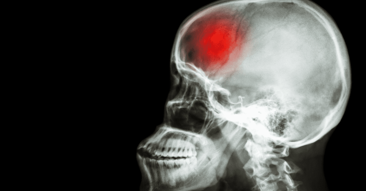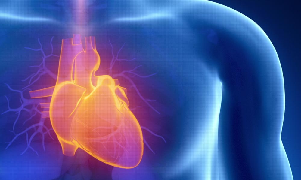
In the past, medical imaging to detect heart disease involved expensive and less accurate procedures. Some methods like treadmill stress testing are not recommended or even dangerous for some patients, particularly those with unstable heart failure, symptomatic severe aortic stenosis, and uncontrolled arrhythmia.
But with the advancement in medical imaging, today’s patients have more options, most of which are safer, more accurate, and even cheaper.
New imaging technologies that can help doctors diagnose heart disease include the following:
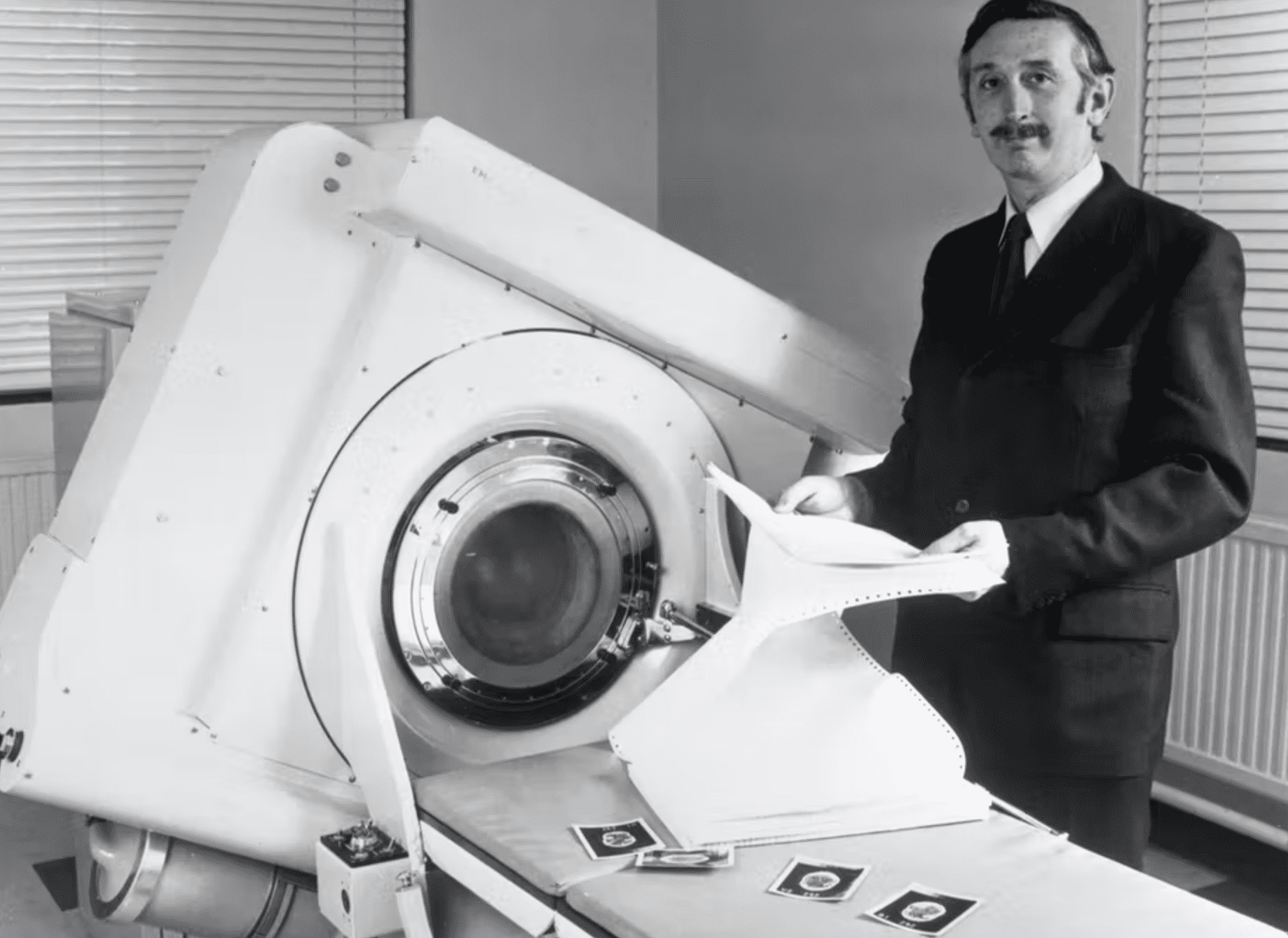
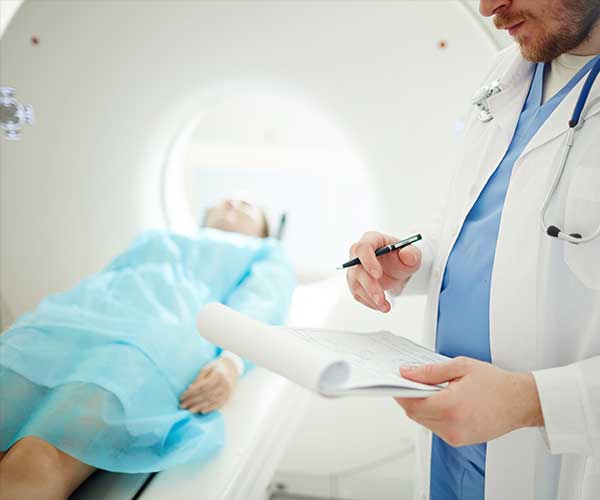
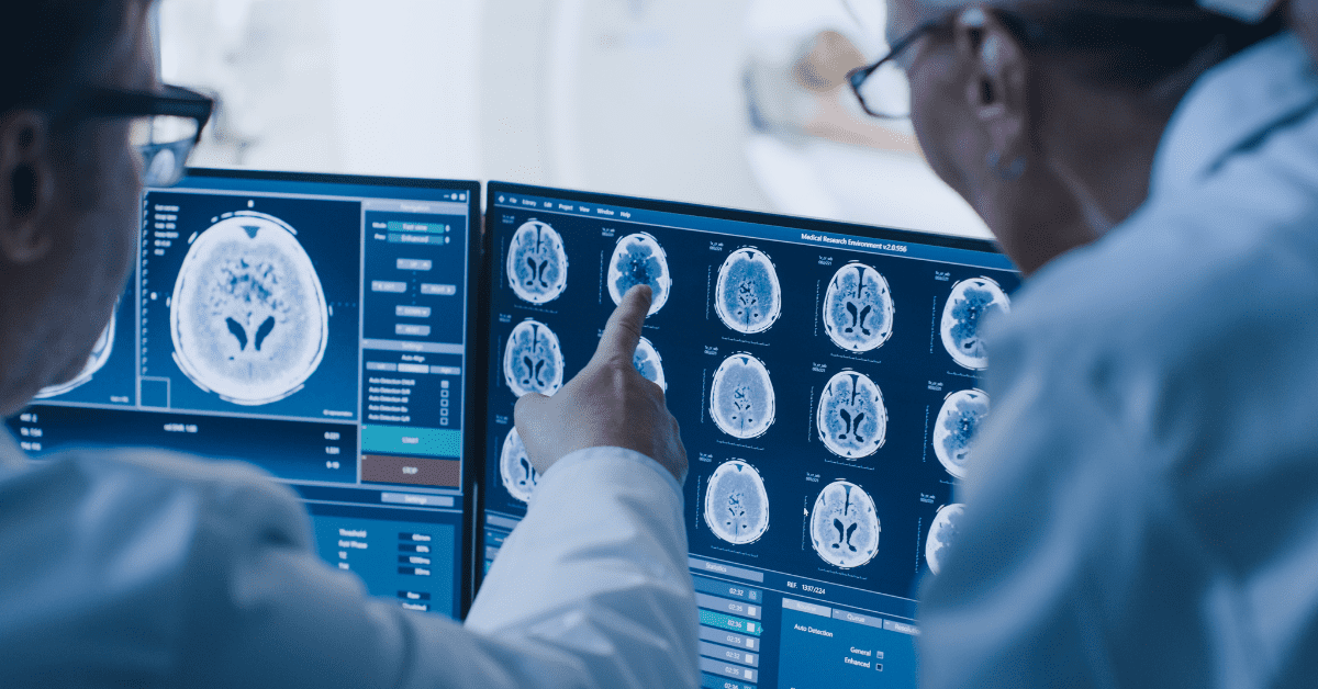
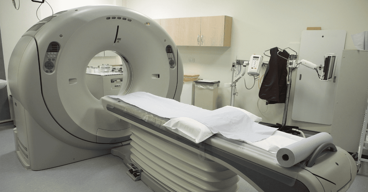
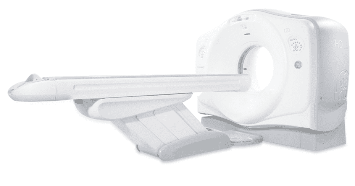
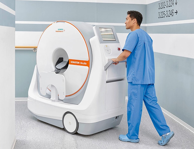
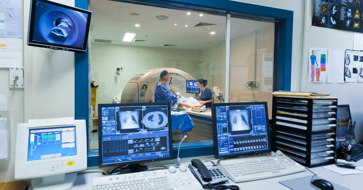
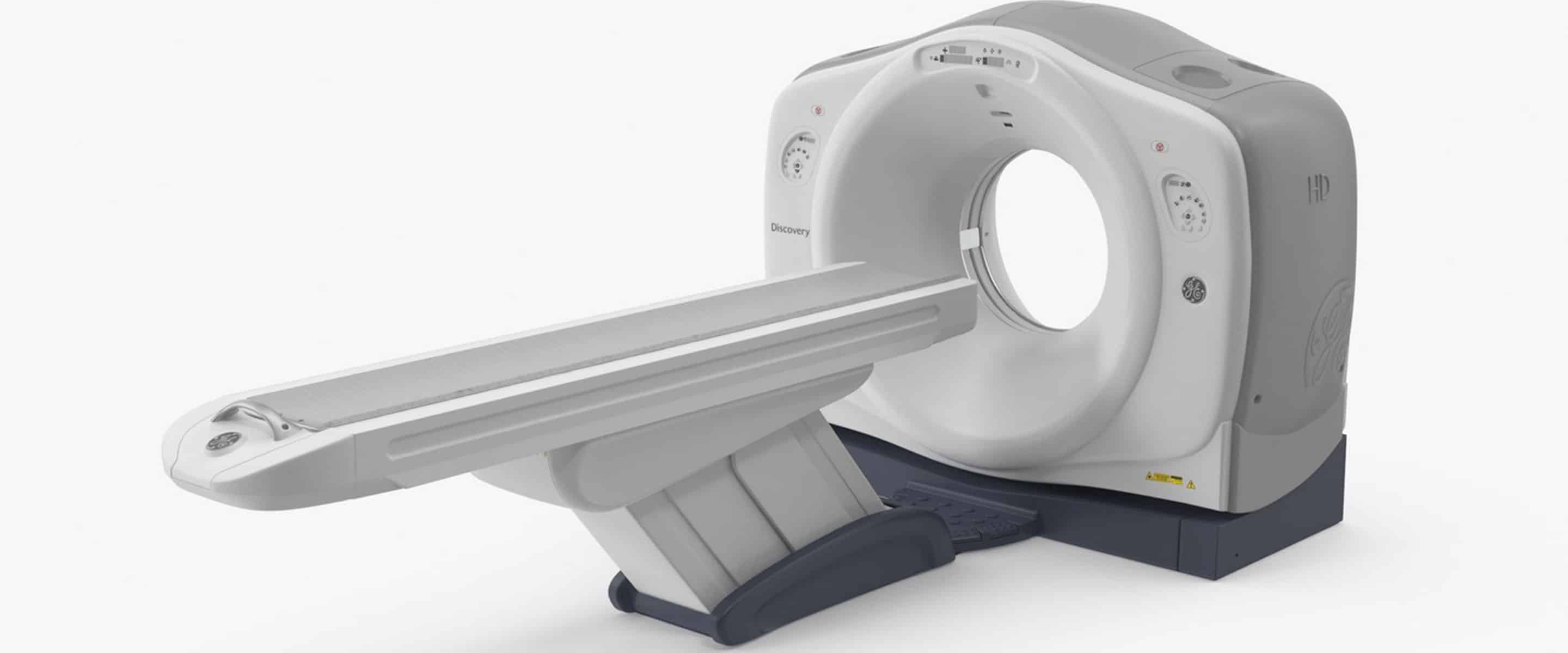
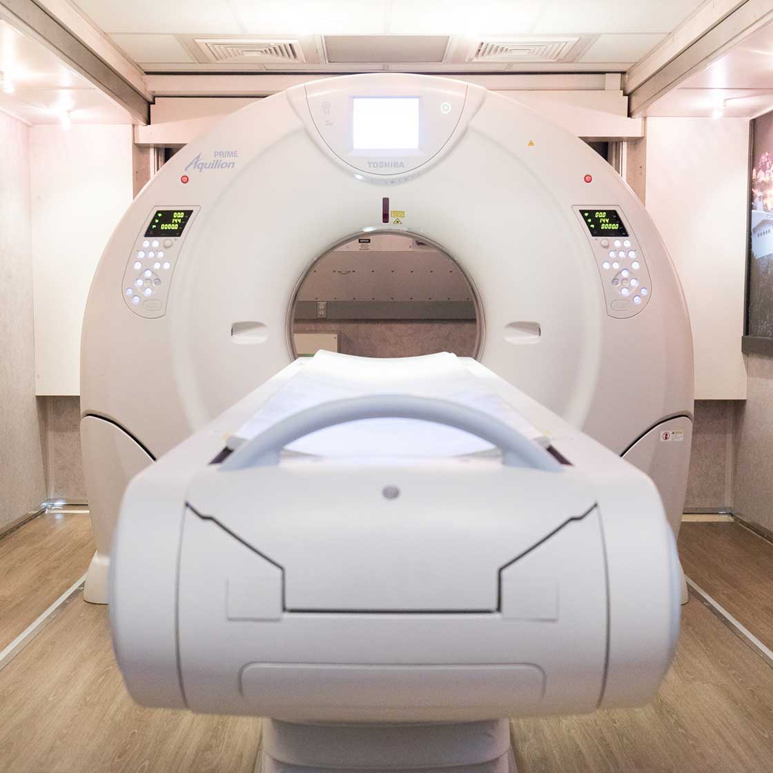
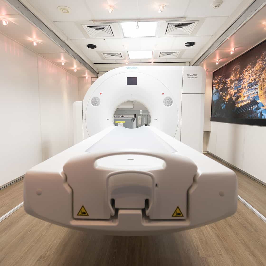
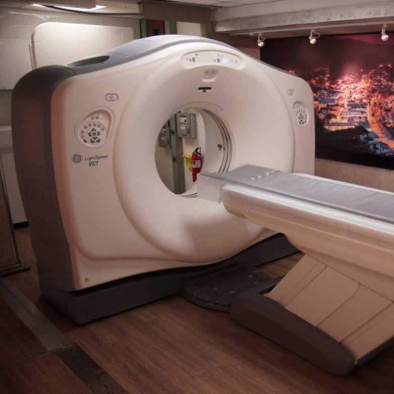
Modern CT scanners can perform a procedure called computed tomography angiography or CTA, which is notable for its accuracy (up to 95%) when detecting small blockages in coronary arteries. The imaging technology also allows doctors to rule out congenital abnormalities and severe heart and artery disease.
CTA is also significantly less invasive than older techniques like the cardiac cath that involves inserting a long, thin tube called catheter into an artery or vein in the neck, groin, or arm to access the heart.
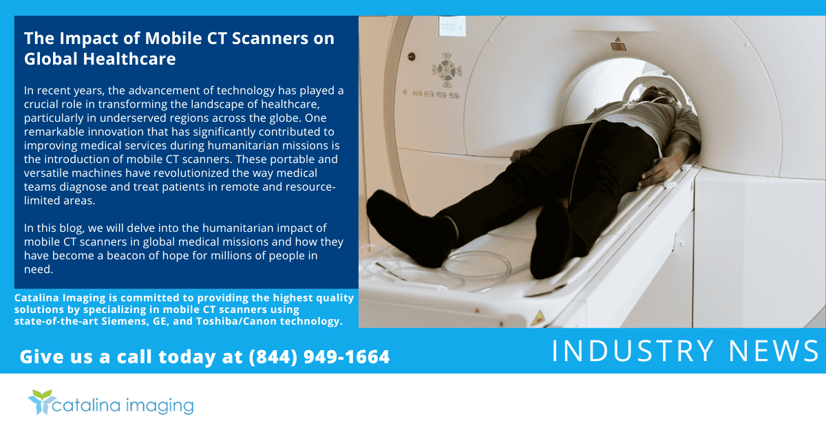
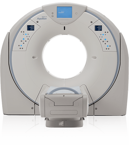
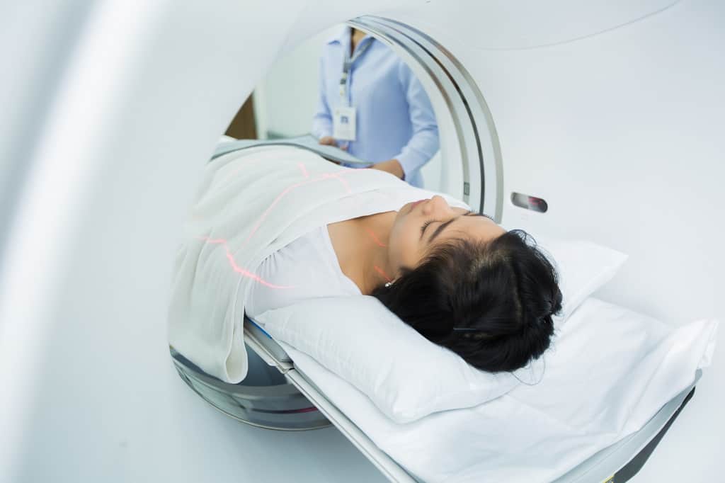
In essence, CT scanners are like X-ray machines that provide better imaging. Before the procedure, a contrast dye is injected into the patient’s arm so the scanner, which looks like a giant donut, can take multiple images to create a more detailed impression of the heart and its blood vessels.
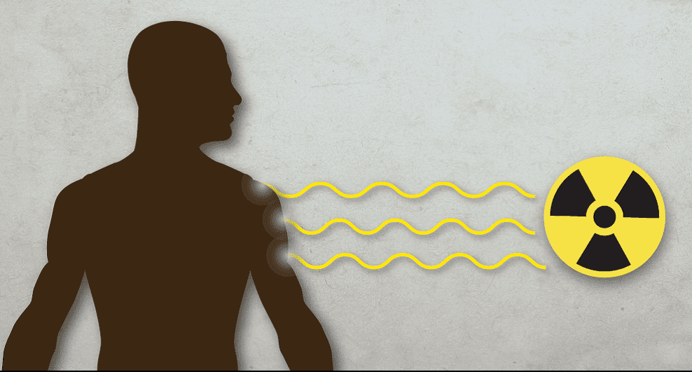
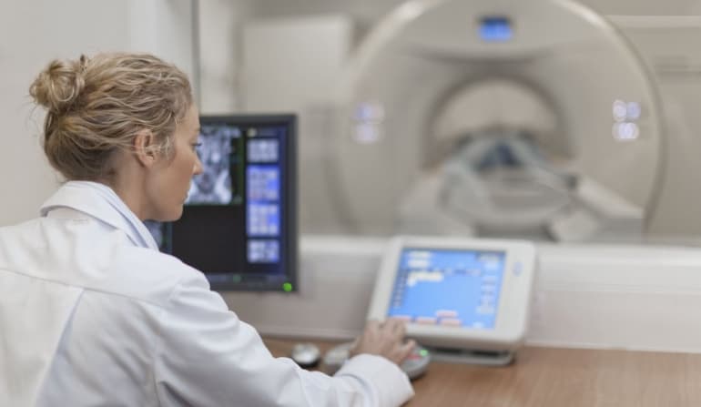
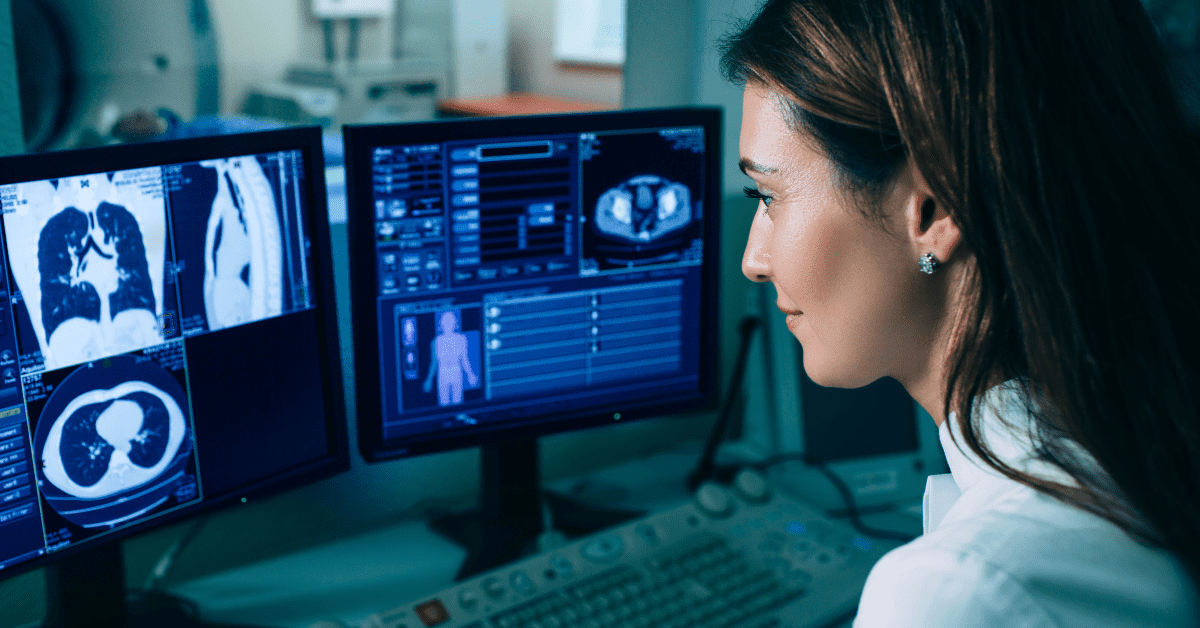
Positron emission tomography or PET scan is an imaging procedure that uses a radioactive substance to detect poor blood flow, buildup of substances in the heart muscle, and other abnormalities in the heart. And when combined with modern CT scanner, the imaging technique can assess not just the heart anatomy but also its function.
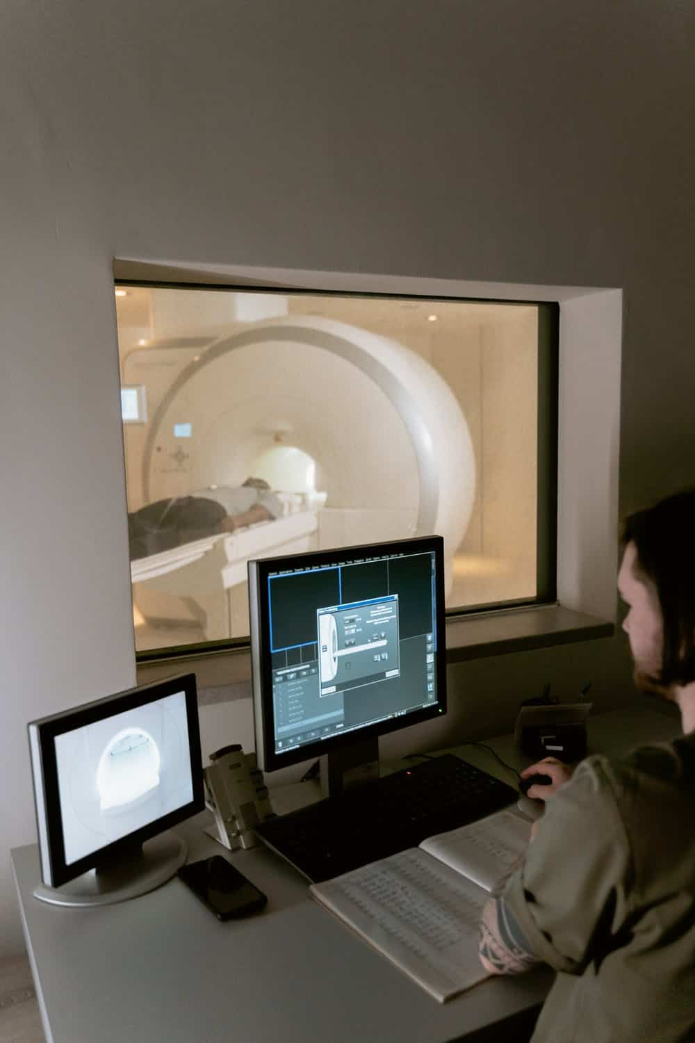
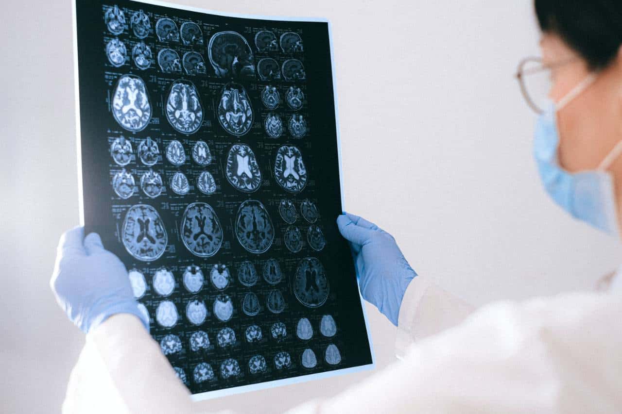
Compared to earlier medical imaging technologies to detect heart diseases, MRI heart scanners provide more detailed images that can help doctors evaluate the heart structure and function.
MRI is particularly useful when it comes to showing valve disease, the heart’s overall appearance (volume, shape, and size), tumors, blood clots, and other abnormalities the older imaging technologies may not detect easily.
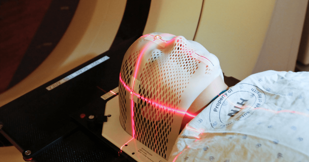
While MRI does not use radiation, it relies on powerful magnets to create detailed images. For this reason, the imaging technique is not performed on patients with a defibrillator or pacemaker.
Nowadays, MRIs come in two versions: open and closed. Open MRI scanners were initially developed to accommodate people with claustrophobia, although they are not an option for medical imaging for heart disease because the organ is in constant motion.
Echocardiography uses high-frequency sound waves called ultrasound to evaluate the valves and heart muscle. Just like in a prenatal ultrasound, it uses a wand-like device that transmits sound waves against the chest area, producing moving images of the heart.
Because the imaging technology does not use any contrast medium or radiation, it can be performed multiple times without predisposing patients to risk of complications.
The list below explains the three types of echo test used today:
The best imaging procedure for the heart boils down to the symptoms, the patient’s medical condition, and the use of adjunct treatments. To learn more about the topic, contact Catalina Imaging at (844) 949-1664 or sale@tshmedical.com.
Catalina Imaging is one of the leading CT mobile imaging providers in the country, serving hospitals and other healthcare facilities in California, Minnesota, North Carolina, and Illinois.