Why Is Mobile Imaging in High Demand Worldwide?
Mobile Imaging is gaining attention worldwide because care often needs to reach patients, not the other way around. In rural clinics, a portable ultrasound can help clinicians assess a pregnancy without a long trip to a distant hospital. In emergency departments, mobile X-ray units can bring imaging to patients who cannot safely move. That matters.
Demand also reflects pressure on crowded facilities. A device that travels between wards may reduce unnecessary transfers and support faster decisions. It does not replace skilled interpretation. Clear images still depend on trained staff, suitable protocols, and careful equipment checks. Small details matter: battery life, image quality, and a stable wireless connection.
Across regions, hospitals, clinics, and home-care services are exploring these systems for different reasons. Some seek quicker bedside assessments; others need imaging options where fixed equipment is scarce. Yet access is uneven. Purchase price, servicing, staff training, and reliable connectivity can limit real-world use. These barriers deserve as much attention as the technology itself.
The term also covers several tools, not one simple solution. Portable ultrasound, digital radiography, and other compact systems serve different clinical needs. Their value depends on the patient, setting, and local workflow. More devices do not automatically mean better care. That is an uncomfortable point.
This article looks at the forces behind global demand, the settings where mobile systems can help, and the practical challenges buyers should weigh. Evidence and local clinical guidance should shape every decision. The promise is real, but it needs careful measurement.
What Mobile Imaging Is and How It Works
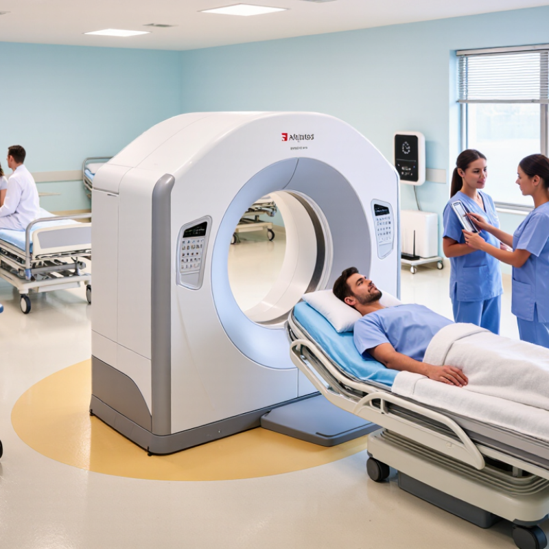
Mobile imaging refers to diagnostic scans performed outside a fixed radiology department, often with portable X-ray or ultrasound equipment. A trained technologist brings the system to a bedside, care facility, or remote clinic. The goal is practical: capture useful images where the patient is, rather than move the patient to the scanner. That matters for people who are frail, isolated, or difficult to transport.
For an X-ray, the technologist positions a detector behind the body part and aims a portable source across it. A brief exposure creates an image that can be reviewed on a screen and sent to a qualified clinician for interpretation. Ultrasound works differently: a handheld probe sends sound waves into tissue and converts returning echoes into moving images. Not magic. Image quality depends on positioning, patient movement, equipment limits, and operator training. Mobile services can reduce transport and waiting, but they do not replace every hospital-based scan. A bedside chest image may answer a focused question, while complex findings can require more detailed imaging. The setting can also be cramped, noisy, or poorly lit; these small details deserve attention.
How Mobile Imaging Technology Has Evolved
Mobile imaging has evolved from bulky, film-based equipment into compact digital systems that can travel to a patient’s bedside. Early portable units often produced limited detail and required careful positioning. Today, digital detectors can capture images quickly and transfer them to clinical systems for review. Lighter components, better batteries, and wireless connections have made imaging more practical in wards, care facilities, and remote settings. The change is more than a smaller machine.
Modern systems can support clearer image previews and faster workflows, while software may help technicians check image quality before a patient leaves the room. Some tools also assist with image analysis, but they do not replace trained professionals or clinical judgment. Network access and data protection remain important, and performance can vary with equipment, connectivity, and staff training. Progress is real, but not perfectly even.
Tips: Keep the detector positioned carefully, confirm patient details, and check image quality before sending the study. If a connection fails, follow the facility’s approved workflow rather than assuming the image was received. A small missed step can create a bigger delay.
Key Factors Driving Global Demand
Mobile imaging is in high demand because care often needs to happen where patients are. A portable X-ray or ultrasound unit can reach a bedside, a rural clinic, or a crowded emergency department. Distance matters. Moving a frail patient across a hospital can be difficult, while bringing equipment to the patient may save time and reduce disruption.
Changing healthcare needs also drive adoption. Aging populations and rising rates of chronic illness create demand for more frequent examinations. Mobile systems can support follow-up visits, home-based care, and screening in locations with limited diagnostic facilities.
In emergencies, clinicians can assess injuries without waiting for a patient to travel to a fixed imaging room. That can help.
Technology has made compact devices more practical, with clearer images and easier digital sharing. Yet a smaller system is not automatically the right choice. Image quality, operator training, equipment maintenance, and secure transfer all affect clinical usefulness.
Not everywhere. A clinic with weak internet or limited technical support may struggle to use connected tools consistently. Even so, mobile imaging can widen access when it is matched to local needs and supported by trained staff.
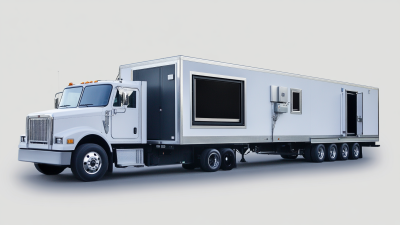
A device in a van, for example, still needs a reliable power source, a clean work area, and a plan for reviewing results.
Where Mobile Imaging Is Used
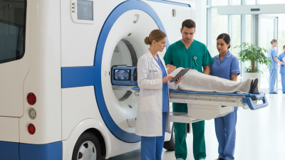
Mobile imaging is used wherever moving a patient is difficult, risky, or likely to delay care. In hospitals, portable X-ray and ultrasound exams can be performed at a bedside, in an emergency department, or in an intensive care unit. A clinician can check a chest image without taking a patient with breathing problems across the building. That matters. Nursing homes and smaller clinics may also use mobile imaging when residents cannot travel easily or local equipment is limited.
Mobile services can reach rural communities, temporary health centers, and disaster response sites. In these settings, imaging may help clinicians assess injuries or guide decisions about referral. It is also used in operating rooms, where images can support procedures while the patient remains in place. Mobile systems are not right for every exam; image quality, room conditions, and access to trained staff still matter. Convenience alone is not enough.
Tips: Before an exam, clear a safe path to the patient and share relevant clinical details with the imaging team. Ask how results will reach the treating clinician. Small delays still happen, especially in crowded wards, so plan for them.
Why Is Mobile Imaging in High Demand Worldwide?
Where Mobile Imaging Is Used
Mobile imaging is used at the bedside in hospitals, in emergency departments and intensive care units, in ambulances, and in rural, remote, and disaster-response settings.
This population split provides context for the need to bring diagnostic imaging closer to patients, particularly in remote areas. It shows where people live, not mobile-imaging usage rates.
Source: World Bank, urban population (% of total), 2023; rural share calculated as the remainder.
Benefits and Challenges of Mobile Imaging Adoption
Mobile imaging brings diagnostic equipment to the bedside, clinic, or remote care site. For patients who cannot easily travel, a portable X-ray or ultrasound exam can reduce transfers and waiting. Less movement. Faster decisions. In a crowded ward, clinicians may get useful images without sending a patient across the building. This can support timely assessment, but mobile exams do not replace every scan or specialist review.
Adoption also brings practical challenges. Image quality can vary with positioning, equipment, and the operator’s experience. Convenience can hide a weak image. Staff need training, and portable X-ray use still requires careful radiation protection. Teams must also keep probes clean, batteries charged, and images securely available to the people involved in care. Poor connectivity can slow that process. These details are easy to underestimate; even a well-equipped service may need time to fit mobile imaging into daily routines.