In the rapidly evolving landscape of healthcare, the role of CT imaging has become increasingly pivotal in enhancing diagnostic accuracy. According to a report by the American College of Radiology, nearly 85 million CT scans are performed annually in the United States, underscoring its critical function in disease detection and management. With advancements in technology, including iterative reconstruction techniques and dual-energy CT, the precision of imaging has seen significant improvements, enabling healthcare professionals to make informed decisions with greater confidence. Moreover, a study published in the Journal of the American Medical Association indicates that adopting advanced CT imaging methods can reduce unnecessary exploratory surgeries by up to 30%. As the medical community continues to harness these techniques, mastering CT imaging will be essential for healthcare providers aiming to enhance patient outcomes and streamline the diagnostic process.
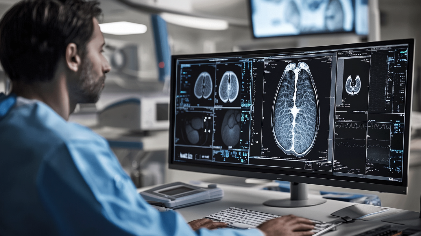
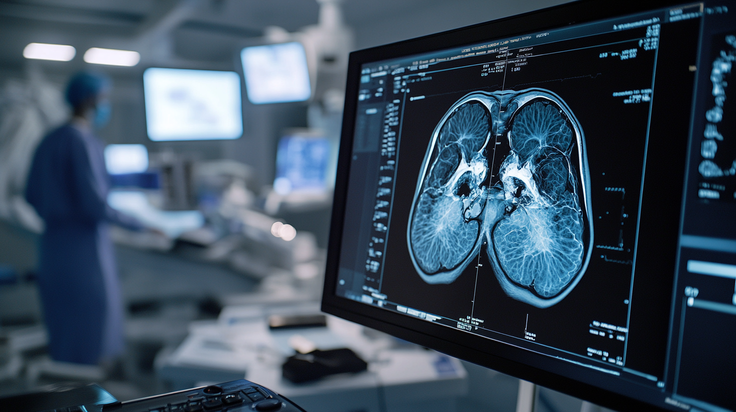
Computed Tomography (CT) imaging has revolutionized diagnostic practices in modern healthcare, providing rapid and detailed insights into a patient’s condition. According to a report by the Radiological Society of North America (RSNA), CT scans account for approximately 73 million procedures performed annually in the United States. This technology employs X-ray equipment to create cross-sectional images of the body, allowing for a comprehensive view of internal structures, which is crucial for accurate diagnosis and treatment planning.
Understanding the fundamentals of CT imaging involves recognizing its advanced capabilities and its impact on patient care. A study published in the Journal of the American College of Radiology noted that the use of CT has significantly improved diagnostic accuracy in conditions such as stroke and cancer detection, with sensitivity rates exceeding 90%. Moreover, as technology continues to advance, newer CT modalities employing iterative reconstruction techniques have been shown to reduce radiation doses by up to 60%, minimizing risks while preserving image quality. As healthcare practitioners refine their skills in CT imaging, the potential for enhanced patient outcomes continues to grow significantly.
In recent years, advancements in CT imaging techniques have significantly enhanced image quality and diagnostic precision in healthcare. Innovative methods such as iterative reconstruction and dual-energy CT have revolutionized the way radiologists interpret scans. Iterative reconstruction allows for the reduction of noise and artefacts, producing clearer images at lower radiation doses. This is particularly crucial in pediatric imaging, where minimizing radiation exposure is a primary concern. By improving the signal-to-noise ratio, these techniques allow for more accurate assessments of complex conditions.
Moreover, dual-energy CT provides a unique advantage by enabling the differentiation of materials based on their atomic number. This technique enhances the visualization of pathological structures and can assist in characterizing lesions more precisely. For instance, it can distinguish between different types of kidney stones or identify the composition of tumors, aiding in the selection of the most effective treatment plan. As these innovative CT techniques continue to evolve, healthcare professionals are better equipped to provide accurate diagnoses, leading to improved patient outcomes and optimized care pathways.
| Technique | Description | Benefits | Common Applications |
|---|---|---|---|
| Dual-Energy CT | Utilizes two different energy levels to enhance material differentiation. | Improved tissue characterization and reduced artifacts. | Oncology, vascular imaging. |
| Iterative Reconstruction | Mathematical algorithms to improve image quality from raw data. | Reduced noise and improved low-contrast detectability. | Body imaging, pediatric patients. |
| Cardiac CT | High-resolution imaging of the heart and coronary arteries. | Non-invasive assessment of coronary artery disease. | Cardiology, pre-surgical evaluation. |
| Cone Beam CT | Uses a cone-shaped X-ray beam for rapid imaging. | Reduced radiation dose and faster scans. | Dental imaging, maxillofacial surgeries. |
| AI-Assisted CT | Integration of artificial intelligence in image analysis. | Enhanced lesion detection and workflow efficiency. | Radiology, oncology. |
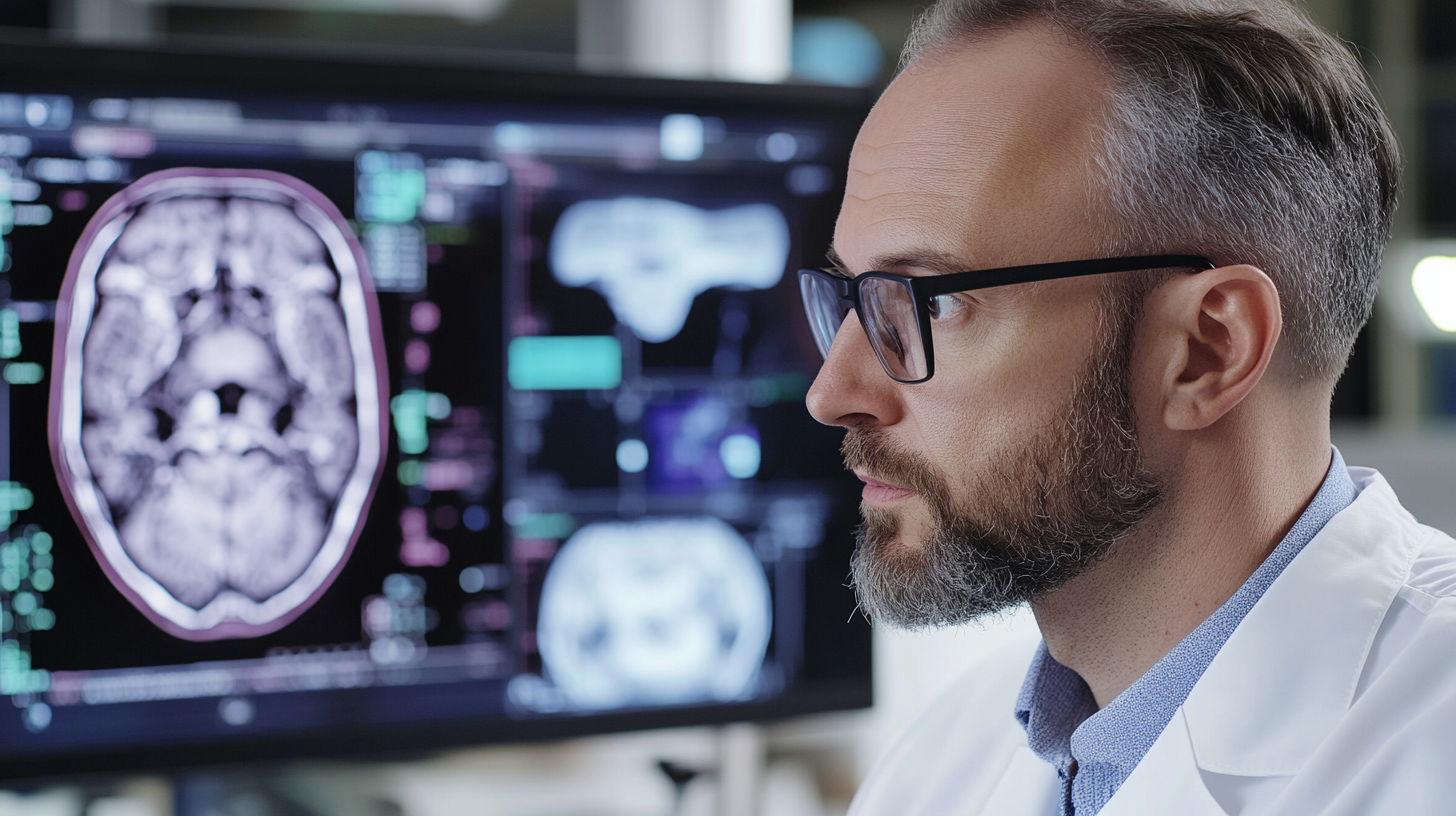
Artificial Intelligence (AI) is revolutionizing the field of CT imaging, enhancing diagnostic accuracy and streamlining imaging processes. According to a report by the American College of Radiology, AI algorithms have the potential to increase the detection rate of critical conditions, such as lung cancer, by up to 15%. By analyzing vast datasets, AI can help radiologists discern subtle patterns and anomalies that may go unnoticed in traditional imaging reviews.
To optimize CT imaging processes, healthcare providers can adopt AI tools that automate routine tasks such as image reconstruction and artifact reduction. This not only accelerates the workflow but also reduces the likelihood of human error. For instance, using AI-driven software for noise reduction can improve image quality without increasing radiation exposure, as highlighted in the Journal of Medical Imaging.
Tips for integrating AI in CT imaging include investing in training programs for radiologists to familiarize them with AI tools, ensuring that the technology is user-friendly and providers can trust its outputs. Additionally, establishing a feedback loop where radiologists can share insights on AI performance can help refine these systems further, ultimately leading to more effective patient care.
CT imaging has become an indispensable tool in modern healthcare, offering unparalleled precision in diagnosing a range of medical conditions. A comparative analysis reveals that CT scans provide higher sensitivity and specificity compared to traditional diagnostic modalities, such as X-rays and ultrasounds. According to a report by the Radiological Society of North America (RSNA), CT imaging can detect subtle pathologies with over 95% accuracy, making it the preferred choice for evaluating complex conditions like trauma, tumors, and vascular diseases.
Moreover, the advantages of CT imaging are underscored by the increasing adoption of this technology in clinical settings. The American College of Radiology (ACR) reported that CT scans account for approximately 10% of all imaging studies but contribute to nearly 50% of the total radiation exposure in patients. This disparity emphasizes the importance of selecting the appropriate diagnostic tool on a case-by-case basis. While MRI and ultrasound present viable alternatives for soft tissue evaluations and pediatric populations, CT imaging remains unmatched in rapid assessments of acute situations. As healthcare continues to evolve, understanding the comparative benefits of CT versus other imaging modalities is crucial for enhancing diagnostic accuracy and patient outcomes.
The future of computed tomography (CT) technology is poised for groundbreaking advancements that promise to significantly enhance diagnostic accuracy in healthcare. Recent reports highlight that the global CT market is expected to reach $8.3 billion by 2026, growing at a CAGR of 5.2% from 2021 to 2026. This growth is primarily fueled by innovations such as iterative reconstruction techniques, which reduce radiation dose while improving image quality. As a result, these advancements not only ensure patient safety but also provide clinicians with clearer, more detailed images for accurate diagnoses.
Additionally, the integration of artificial intelligence (AI) in CT imaging is revolutionizing how radiologists interpret results. A study published in the journal "Radiology" found that AI algorithms could improve detection rates of pulmonary nodules by up to 15%. By augmenting clinical decision-making, AI tools help in prioritizing urgent cases and streamlining workflows in busy medical facilities. These trends not only enhance the clinical implications of CT imaging but also signify a shift towards more precision-driven healthcare. As these technologies continue to evolve, the potential for better patient outcomes and more efficient healthcare delivery becomes increasingly attainable.